The items you find are only part of our catalog. We are in the process of completing the insertion
-
Instruments
- Autoclaves
- Balances (Laboratory and medical)
- Centrifuges (Laboratory)
- Chillers
- Climatic chambers
- Colony counters
- Conductometers
- Cryoscopy
- Data-logger
- Digital laboratory thermometers
- Evaporators - Concentrators
- Freezers (Medical, laboratory and drug)
- Furnaces (Industrial and Lab)
- Heat sealers
- Heating mantles for laboratory
- Heating plates (Laboratory)
- Homogenizers
- Hoods (Laboratory)
- Ice makers
- Incubators and refrigerated thermostats (Laboratory)
- Laboratory pumps
- Loop sterilizers
- Melting point determination tools
- Microscopes
- Multiparameter analyzers
- Ovens (Laboratory)
- Oximeters
- pH-meters
- Polarimeter
- Professional measurement and control tools
- Refractometers
- Refrigerators (Medical, laboratory and drug)
- Safety cabinets
-
Shakers (Laboratory)
- Magnetic heating stirrers
- Magnetic stirrers
- Multi-position heating magnetic stirrers
- Rod agitators
- Vortex shakers for test tubes
- Alternating movement agitators (horizontal plane)
- Rotating stirrers (horizontal plane)
- Tilting movement agitators
- Rotating disc shakers (for tubes)
- Petri dish stirrers
- Rotating rack agitators
- Roller stirrers for hematology
- Shaking water baths
- Spectrophotometers
- Thermal cycler
- Thermoblocks for test tubes and thermoreactors COD
- Thermomixer
- Ultrapure water systems
- Ultrasound laboratory baths
- Viscometers
- Water baths
- Water distillers - demineralizers
- Lab Benches, Hoods and Technical Laboratory Furniture
-
Glassware - Porcelain - Quartz
- Graduated glass pipettes with syringe
- Beaker
- Bottles and caps
- Bottles Pharmaceutical
- Burettes
- Capillary tubes
- Counting chambers
- Crucibles
- Crystallizing dishes
- Dropper Ranvier
- Dryers
- Erlenmeyer flasks
- Evaporating dishes
- Flasks
- Funnels
- Glass syringes
- Graduated cylinders
- Graduated glass pipettes - calibrated
- Incinerating dish
- Jars
- Laboratory bottles
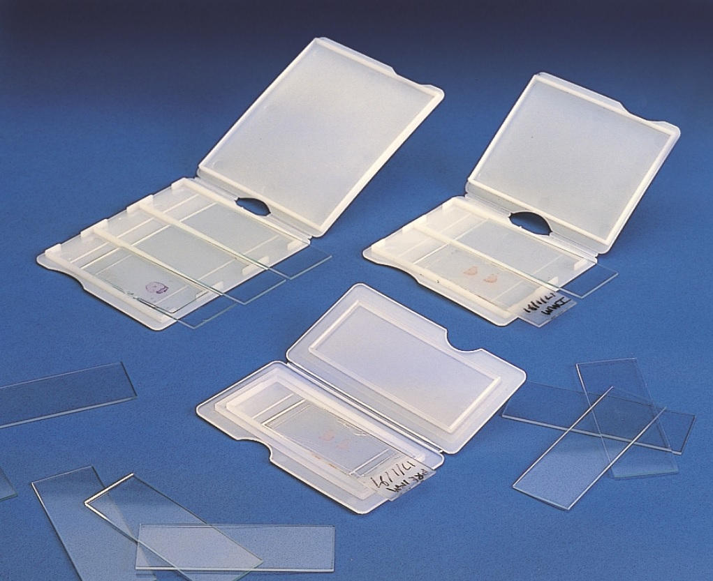
- Microscopy slides
- Mortars and pestles
- Optical glass - quartz cuvettes
- Pasteur glass pipettes
- Petri dishes
- Pycnometers
- Sedimentation cones
- Spheres
- Spirit lamps
- Staining dishes
- Test tubes
- Vacuum flasks
- Various glassware
- Vials
- Viscometers
- Volumetric flasks
- Watch glasses
- Weighing bottles in borosilicate glass
- Wintrobe hematocrit tubes
-
Reusable Plastics
- Basins
- Beaker in plastic
- Bottles and flasks for laboratory in Plastic
- Boxes in plastic
- Containers for 24 h urine collection
- Containers with screw cap
- Cryobox - boxes for cryogenics
- Cylinders in plastic
- Jars for staining slides
- Jugs in plastic
- Magnetic stirrups and rods
- Mini Cooler
- Oilers
- Portable plastic fridge
- PTFE watch glasses
- Rack and cuvette holders
- Slide holder (cases, boxes, trays)
- Soaked in plastic
- Tanks and drums
- Urinals and bedpans for the sick
- Water jet pump
-
Disposable Plastics
- Disposable test tube holder
- Bags
- Blood analysis devices
- Blood group plates
- Caps for test tubes
- Cell culture plastics
- Containers
- Counting chambers in plastic
- Cryovials
- Cups and cuvettes for instrumentation
- Forceps
- Graduated serological pipettes
- Histology - Pathological anatomy
- Inoculation loops and rods for bacteriology
- Microvials
- Pasteur Plastics pipettes
- Plastics Life Sciences
- Plates for micromethods (microtiter)
- Polystyrene Petri dishes
- Reagent trays
- Swabs
- Syringes for microdispensers - dispensing tips
-
Test tubes in plastic
- Disposable plastic tubes with conical bottom
- Disposable round bottom plastic tubes
- Disposable flat bottom plastic tubes
- Conical bottom tubes with screw cap for centrifuge (Falcon type)
- Urine collection tubes
- Microtubes for centrifuge with conical bottom
- Test tubes for serum separation
- Test tubes with anticoagulant
- Pediatric - veterinary sampling tubes
- Tips for micropipettes
- Water sampling bottles
- Weighing boats
-
Tubes and Vacuum Blood Collection Systems
- Accessories for blood sampling
- Set for venous sampling
-
Vacuum collection tubes
- Tubes with separator gel and coagulation activator
- Tubes with separator gel and K2 EDTA
- Dry tubes with coagulation activator
- Tubes with lithium heparin
- Provette with K3 EDTA
- Tubes with KF Na2 EDTA glycolysis inhibitors
- Tubes for coagulation and PTT tests with Na Citrate
- Test tubes for ESR
- Kimased - test tubes and accessories for ESR
- Test tubes without additive
- Urine tubes
- Filtration
-
Liquid Handling
- Bottle dispensers
- Continuous bottle-top burette
- Microdispenser
- Micropipettes for dilution
- Multichannel electronic micropipettes
- Multichannel variable volume micropipettes
- Pipette aspirators
- Pipettors
- Positive displacement micropipettes
- Reagent dispensers
- Single channel electronic micropipettes
- Single-channel variable volume micropipettes
- Lab consumables
-
Lab equipments
- 18-10 stainless steel glasses
- Cart
- Contacellule
- Containers for cryogenics
- Dewar vessels
- Dryers for glassware (Lab)
- Fridge box
- Gas burners - Bunsen lamps
- Istoteche
- Melting point equipment
- Sieves
- Stainless steel racks for freezers
- Stainless steel racks for Petri dishes
- Stopwatches, timers, laboratory minute counters
- UV germicidal lamps for instruments
- Water suction pumps for vacuum
-
Metal clamps and stands
- Baskets in stainless steel
- Forks
- Ball joint pliers
- Clamp for burettes
- Clamps
- corkscrew
- Crucible tongs
- Framework base
- Lift tables
- Manual pliers for closing - opening aluminum ring nuts
- Pipes for sterilization
- Pliers for conical joints
- Rubber hose clamps (Hoffmann, Mohr)
- Slide clamps
- Stainless steel spatulas
- Support pliers
- Support rings
- Supports
- Test tube holder in steel
- Tongs for beakers
- Tongs for capsules
- Tongs for matrasses
- Triangles for crucibles
- Tripod supports
- Pinzeria and surgical instruments
- Safety and protection items
- Sterilization products
- Sanitary items
- Thermometry - Densimetry
- Diagnostics - Microbiology
- Chemical reagents
- Papers - Reactive kits
- Molecular and cellular biology reagents
- Columns and consumables for GC - HPLC
- Disinfectants
- Detergents for laboratory use
Related Products
-
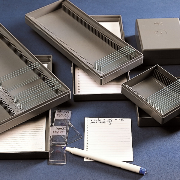
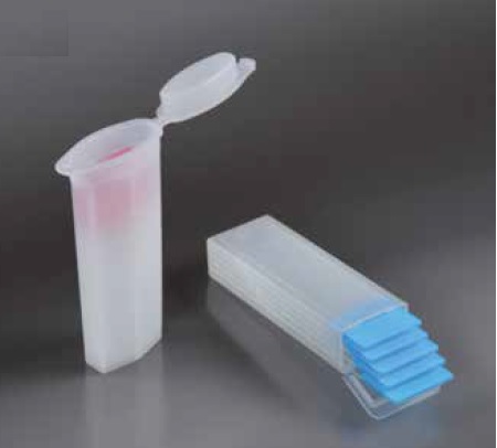
Microscope slide boxes
for 76x26 mm slides -
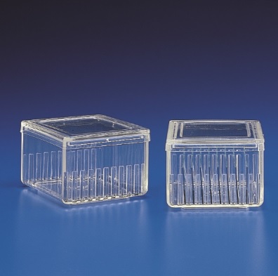
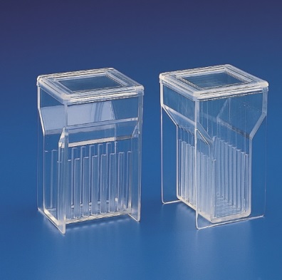
Staining jar Schifferdecker
-
Staining jar with 2 lids in PMP
-
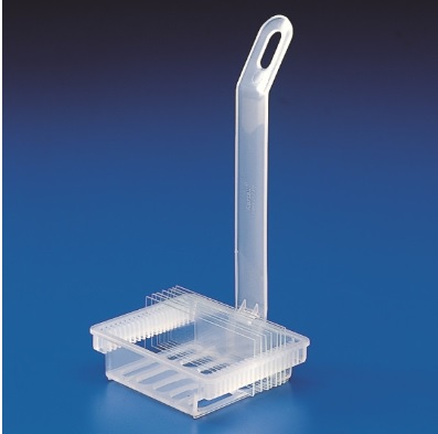
Staining rack
-
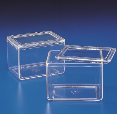
Staining jar Hellendhal
-
Tray for microscope slides
20 places -
Tray for microscope slides
40 places -
Tray for microscope slides
-
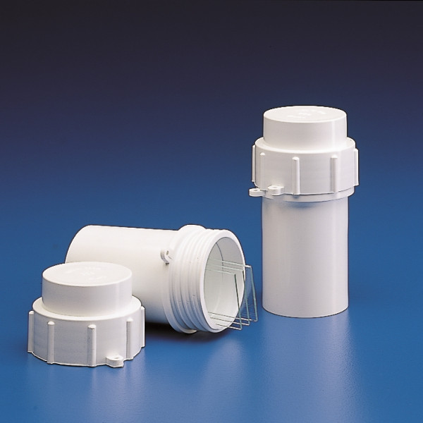
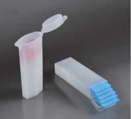
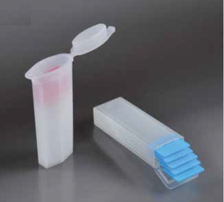
Cylindrical postal slide holders
-
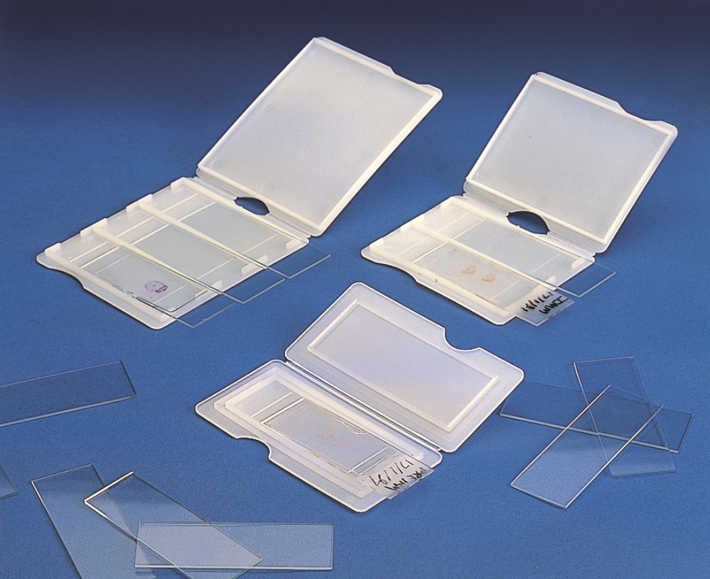
Postal slide holder in PE
1 place -
Postal slide holder in PE
3 places -
Postal slide holder in PE
2 places -
Postal slide holder in PE
-
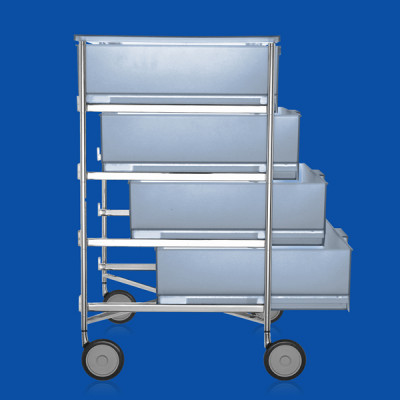
Four-drawer cabinet with swivel wheels
-
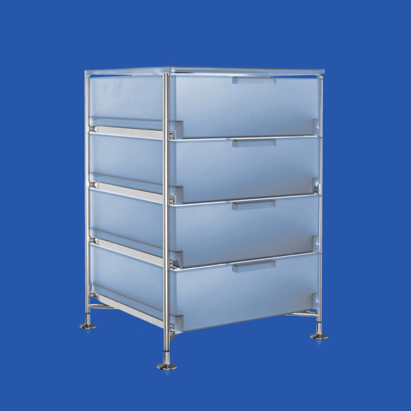
Cabinet with four drawers with support feet
-
Postal slide holders
-
Postal slide holders
-
Postal slide holders
-
Indelible laboratory marker fine tip black color
-
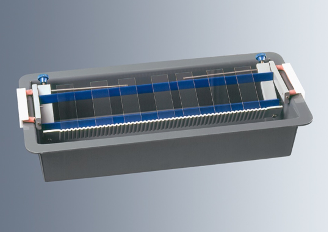
Staining bridges
-
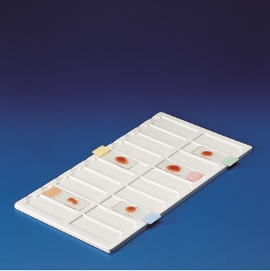
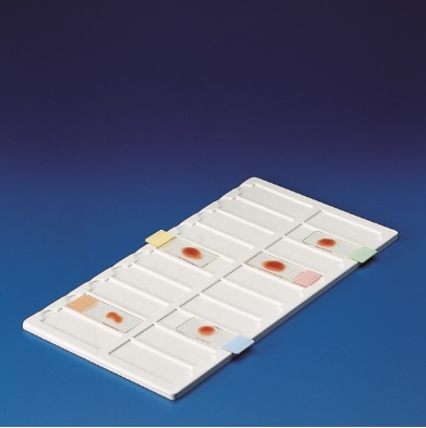
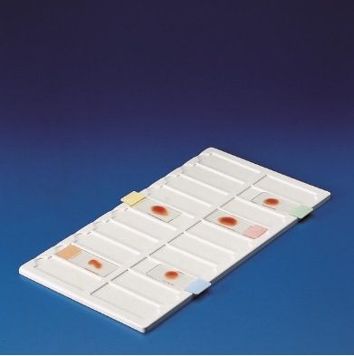
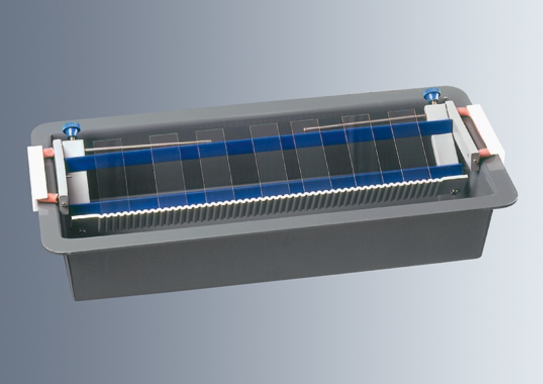
Staining trays
CONTACTS
ENRICO BRUNO s.r.l.
Via Duino 140
10127 Torino Italy
Tel. +39 011 3160808
Fax +39 011 3160753
PEC pec@pec.enrico-bruno.it
CF/P.IVA: 04891320014
WRITE TO US
For orders, prices requests and offers, technical service commerciale@enrico-bruno.it
For administration service amministrazione@enrico-bruno.it
For Direction – Purchase Department acquisti@enrico-bruno.it
